What is 3D & 4D Ultrasound Scan?
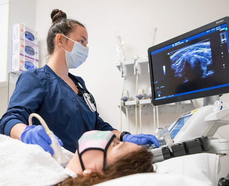
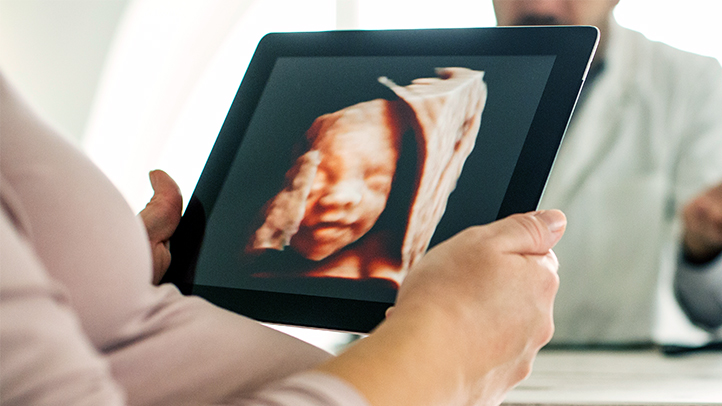
3D imaging allows for the visualization of fetal structures and the internal anatomy as static 3D images.
4D ultrasounds allow for a live-streaming video of the images, showing the motion of the fetal heart wall or valves, as well as the current blood that is flowing through various vessels.
- Antenatal Ultrasound
- General Ultrasound
- Women Imaging
- Musuloskeletal Ultrasound
- Paediatric Ultrasound
- Doppler Studies
- USG Guided Intervention
- Prenatal Screening
- Adult Echo Cardiogram (ECHO)
- ElectroCardiogram (ECG)
WHO NEEDS IT
Who Need 3D & 4D Ultrasound?
3D ultrasound is mainly useful for pregnancy people to examine the fetal heart rate in real-time. Among other things, it is also useful for facilitating the characterization of some congenital defects, such as skeletal anomalies and heart issues. With real-time 3D ultrasound, the fetal heart rate can be examined in real-time.
Antenatal Ultrasound
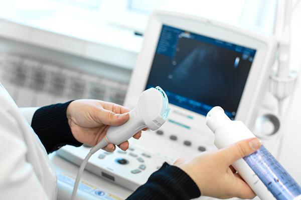
This ultrasound can confirm your pregnancy is viable and estimate your baby's due date. It can also confirm how many babies you are carrying and check that your baby is growing in your uterus, and is not ectopic (growing outside the uterus).
- Early Pregnancy Scan
- Nuchal Translucency(NT) Scan
- (11-13 weeks 6 days)
- Anomaly/TIFFA Scan
- (18-20 weeks)
- Growth Scan
- (26 weeks onwards)
- Biophysical Profile and doppler
- (28 weeks onwards)
- Foetal echo
- (16-22 weeks)
General Ultrasound
Ultrasound provides real-time imaging. This makes it a good tool for guiding minimally invasive procedures such as needle biopsies and fluid aspiration.
- Abdomen & Pelvis / KUB
- Thorax
- Scrotum
- Neck / Thyroid
Women Imaging
Women's imaging covers several diagnostic imaging procedures that specifically apply to women and determine diseases that are most common in females, such as gynecological complications or breast cancer.
- Ultrasound Pelvis (TAS/TVS)
- Follicular Scan
- Infertility work-up imaging
- Ultrasound breast (Sonomammography)
Musuloskeletal Ultrasound
Musuloskeletal ultrasound imaging uses sound waves to produce pictures of muscles, tendons, ligaments, nerves and joints throughout the body. It is used to help diagnose sprains, strains, tears, trapped nerves, arthritis and other musculoskeletal conditions.
- High resolution sonography of joints - Shoulder / Knee / Ankle / Wrist / Finger High Resolution Soft Tissue Scan


Paediatric Ultrasound
Paediatric ultrasound helps in imaging the infectious diseases which are common in Paediatric patients that involves no irradiation or sedation and can be performed repeatedly at the patient's bedside.
- Neurosonogram
- Paediatric / Neonatal hip
- Neonatal Spine
Doppler Studies
Doppler ultrasound is a noninvasive test that can be used to measure the blood flow through your blood vessels. It works by bouncing high-frequency sound waves off red blood cells that are circulating in the bloodstream.
- Arterial Lower / Upperlimb Doppler
- Venous Doppler for DVT / Varicose Venis
- Renal Arterial Doppler
- Carotid & Vertebral (4 vessel) Doppler
- AV Fistula mapping
- (Pre & Post dialysis)
- Abdominal Doppler
- (Aorta / Portal Venous)


USG Guided Intervention
USG Guided Intervention is diagnostic or therapeutic minimally invasive procedures guided by real-time ultrasound imaging, preferably using attachable needle steering devices to locate and examine fluid collection in tendons, muscles, cysts and soft-tissue masses, so that: Fluid can be accurately extracted.
- FNAC
- Biopsy
- Drainage
Prenatal Screening
Prenatal Screening can be used to detect some birth defects at various stages prior to birth. prenatal screening is prenatal care that focus on detecting problems with the pregnancy as early as possible.
- Down's Screening
- Triple Marker Test
- Quadruple Marker Test


ECG
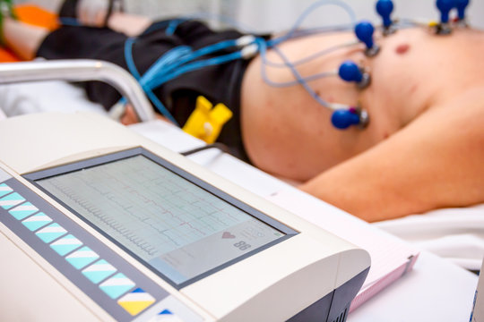
An electrocardiogram (ECG) is a painless test that records the electrical signals in the heart. It is commonly used to quickly detect heart problems and monitor the heart's health.
ECHO
An echocardiogram uses ultrasound to check for anomalies in the heart's structure.

Advantages of 3D & 4D Ultrasound
Non-invasive
Ultrasound scans are non-invasive, which eliminates the need for surgery.
Accurate visualization
Provides detailed images inside the body which helps for diagnosis.
Reduces multiple integrations
No need of multiple integration of images as in 2D.

